Comparative Physiological Aspects Of Vertebrates
Reproduction is the process by which plants and animals produce offspring.
Introduction
Much of biology is based on the principles of chemistry. cells are composed of organic molecules, so a discussion of cells would not be complete without talking about these substances. Chemistry is also important in that organisms perform chemical reactions to store and release the energy that they use to carry out their various processes, and the way cell organelles work has a simple basis in chemistry.
Inside of every cell are tiny structures which scientists refer to as organelles. These organelles, which carry out all sorts of processes, including respiration, digestion, excretion, reproduction and many others. We'll focus on the different types of organelles, what the function of each one is, and how the principles of chemistry allow it to perform this function.
Although cells are remarkable for the complexity of the structures which they contain, even more incredible are the great many processes which occur in cells. Like multicellular organisms, cells also have a way of eating, breathing, and reproducing. This review focuses on how cells reproduce and forms new offsprings.
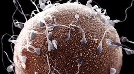
One of the requirements which something must satisfy to be considered alive is that it must reproduce. Animals and plants, generally have very complex methods and patterns of reproduction. Instead of utilizing sexual reproduction like animals (which involves two parents of different sexes), most unicellular organisms undergo a process of asexual reproduction, whereby only one parent cell is needed in the reproductive process. This review is devoted to explaining this important process. The beginning of life is a single cell that results from the union of a haploid (n) egg and a haploid (n) sperm.
Life origin
The speculations about the origin of life were difficult due to less details of the chemical basis of life. Darwin (1809-1882) could express by doubtfully speculation about the bounding between non-chemical substances and life as "in some warm pond with all kinds of ammonia and phosphoric salts, light, heat and electricity" might have been the origin of life. Louise pasteur (1822-1895) began to test how easy it is to make the transition from non-living chemical systems to microbial life,but they had no understanding of the differences between environmental conditions in the current biosphere of earth and conditions when life first arose.
Pouchet (1800-1872) claimed that boiled water and straws provided suitable conditions for the generation of new microbes. Pasteur showed that pouchet's methods allowed living microbes back into the boiled straws after cooling. Reproduction is an important for the survival of all living things. Without reproduction, life would come to an end. In other ward, reproduction is biological process by which new individuals are produced from their parents. Also, Reproduction is a fundamental event of all known life where each individual organism exists as a result of reproduction. Some animals do not reach sexual maturity for many years after birth and even then produce few offspring. Others reproduce quickly, most offspring do not survive to adulthood. Animals with few offspring can devote more resources to nurturing and protection of each individual offspring, thus reducing the need for many offspring. On the other hand, animals with many offspring may devote fewer resources to each individual offspring; for these types of animals it is common for many offspring to die soon after birth, but enough individuals typically survive to maintain the population.
Life without reproduction
The existence of life without reproduction is the subject of some speculation. The biological study of how the origin of life led from non-reproducing elements to reproducing organisms is called abiogenesis.
Several scientists have succeeded in producing simple viruses from entirely non-living materials. The virus is often regarded as not alive. Being nothing more than a bit of RNA or DNA in a protein capsule, they have no metabolism and can only replicate with the assistance of a hijacked cell's metabolic machinery.
Reproduction is the biological process by which new individual organisms are produced from their parents. Reproduction is a fundamental feature of all known life; each individual organism exists as the result of reproduction.
Forms of reproduction
Reproduction By Pollination
Pollen is a fine, powdery substance consisting of microscopic grains containing the male gametophyte of certain plants that reproduce sexually. These plants include angiosperms, a type of plant that produces flowers during sexual reproduction, and gymnosperms, which reproduce sexually through the use of seeds that are exposed and not hidden in an ovary, as with an angiosperm. Pollen is designed for long-distance dispersal from the parent plant, so that fertilization can occur. Pollination is the transfer of pollen from the male reproductive organs to the female reproductive organs of a plant, and it precedes fertilization. In other words, pollination is the equivalent of sexual intercourse for seed-bearing plants. Actually, cross-pollination, or the transfer of pollen from one plant to another, would perhaps be analogous to sexual intercourse in animals. Pollination occurs in seed-bearing plants, as opposed to the more primitive spore-producing plants, such as ferns and mosses. Gymnosperms, such as pines, firs, and spruces, produce male and female cones, whereas angiosperms produce flowers containing a male organ called the stamen and a female organ called the pistil. Both types of plants depend on insects and other creatures to aid in the pollen transfer.
Allogamy
Allogamy is a biological reproduction of an ovum from one individual with the spermatozoa
of another.
Autogamy
It occurs in hermaphroditic organisms where the two gametes fused in fertilization come from the same individual.
Production Of Somatic and Genital Cells
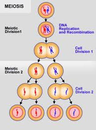
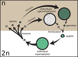
Cell reproduction involves one of two pathways. All cells except for those sex cells producing gametes go through the cell cycle and mitosis. Cells destined to become gametes (sperm and eggs) also go through the cell cycle. However, instead of dividing up the genetic material via the process of mitosis, which retains the same amount of DNA in each daughter cell, they undergo meiosis. Mitosis produces two diploid daughter cells and meiosis produces four haploid daughter cells .
Mitosis: Mitosis is the cell division process in which a eukaryotic cell divides the chromosomes into two identical sets of two daughter nuclei in its cell nucleus. It is followed by cytokinesis, which equally divides the nuclei, cytoplasm, organelles and cell membrane into two daughter cells. Mitosis and cytokinesis together form the mitotic (M) phase of the cell cycle. The sequence of events are divided into different stages named as prophase, prometaphase, metaphase, anaphase and telophase.
-Phases Of Mitosis-
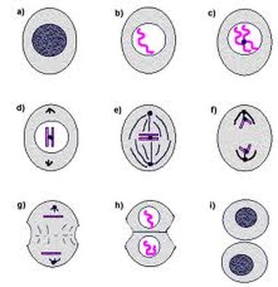
Mitosis occurs in different ways in different species. For example, animals undergo an open mitosis process in which the nuclear envelope breaks down before the chromosomes separate, while fungi and yeast undergo a closed mitosis in which the chromosomes divide within an intact cell nucleus.
-Phases Of Meiosis-
Meiosis
It is a process of reductional division in which the number of chromosomes per cell is halved. Before it begins, the DNA in the original cell is duplicated during S-phase of the cell cycle. Meiosis separates the replicated chromosomes into four haploid gametes or spores. If it produces gametes, these cells should fuse during fertilization to create a new diploid cell or zygote. In plants, meiosis produces spores which results in the formation of haploid cells that can divide vegetatively without undergoing fertilization. The different stages involved in meiosis are meiosis I, prophase I, metaphase I, anaphase I, telophase I and meiosis II. Meiosis is necessary for sexual reproduction and therefore occurs in all eukaryotes that reproduce sexually. Meiosis does not occur in archaea or bacteria as they reproduce asexually through binary fission process.
Difference Between Mitosis and Meiosis
Mitosis is the cell division process that occurs in our body and does not change the number of chromosomes in the cell. It happens in all somatic cells of the body and helps to make new cells. In meiosis, the number of chromosomes is halved and therefore, it is also known as Haploid Division. It helps in variation in the offspring and sex determination, as half the chromosomes are from the father and half from the mother.
Table: The differences between mitosis and meiosis
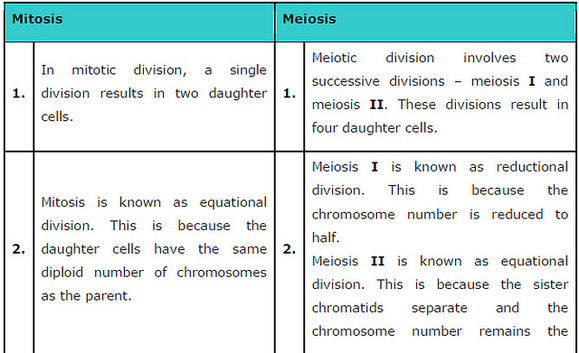
The main difference between mitosis and meiosis are
illustrated as in the following table.
Alternative Between Sexual and Asexual Reproduction
Some species alternate between the sexual and asexual ways, an ability known as heterogamy, depending on conditions. For example, the freshwater crustacean Daphnia reproduces by parthenogenesis in the spring to rapidly populate ponds, then to sexual as the intensity of competition and predation increases.
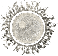
Fertilization
The process by which two gametes fuse to become a zygote, which develops into a new organism. The resultant zygote is diploid. In cross-fertilization, the two gametes come from two different individual organisms. In self-fertilization, the gametes come from the same individual. Fertilization includes the union of the cytoplasm of the gametes (called plasmogamy) followed by the union of the nuclei of the two gametes (called karyogamy). Among many animals, such as mammals, fertilization occurs inside the body of the female. Among fish, eggs are fertilized in the water. Among plants, fertilization of eggs occurs within the reproductive structures of the parent plant, such as the
ovules of gymnosperms and angiosperms. The reproduction is subdivided according to types of fertilization into three main types.
(a) Internal fertilisation
During internal fertilisation, eggs are fertilised inside the female's body. Animals, like reptiles and birds, lay eggs after fertilisation. New offspring develop outside the female's body. All eggs are covered by a protective shell except mammal females, in which a new embryo develop inside their
body.
(b) External fertilisation
During external fertilisation, the egg is fertilised outside the female's body. Male
and female gametes are released into these species' surroundings where they
fuse, forming a zygote. This type of fertilisation usually occurs in water as
in amphibians and fish.
(c) Hermaphrodites
Hermaphrodites are animals that have both female and male reproductive organs. Earthworms and leeches are hermaphrodites, but as they produce eggs and sperm at different times, they need a mate to reproduce. Flatworms are hermaphrodites that can self-fertilise.
All living things reproduce. Reproduction is the process of generating offspring. There are two main types of reproduction: sexual and asexual. Some organisms reproduce by only one type of reproduction and others can reproduce by both.
Sexual reproduction
Sexual reproduction is the process of producing offspring for the survival of the species, and passing on hereditary traits from one generation to the next. The male and female reproductive systems contribute to the events leading to fertilization. Then, the female organs assume responsibility for the developing embryo, birth, and nursing. The male and female gonads (testes and ovaries) produce sex cells (ova and sperm) and the hormones necessary for the proper development, maintenance, and functioning of the organs of reproduction and other organs and tissues.
Even some groups of organisms that practice asexual reproduction such as fungi, certain protists and vascular plants, various invertebrates and even some reptiles and amphibians exhibit sexual reproduction as well.
Asexual reproduction
It is the process in which an organism produces identical copy of itself without a contribution of genetic material from another individual. Asexual reproduction is the primary form of reproduction for single-celled organisms such as the archaea, bacteria, and protists. Many plants and fungi reproduce asexually as well. While all prokaryotes reproduce asexually (without the formation and fusion of gametes), mechanisms for lateral gene transfer such as conjugation transformation and transduction are sometimes likened to sexual reproduction. Asexual reproduction includes numerous types as:
illustrated as in the following table.
Alternative Between Sexual and Asexual Reproduction
Some species alternate between the sexual and asexual ways, an ability known as heterogamy, depending on conditions. For example, the freshwater crustacean Daphnia reproduces by parthenogenesis in the spring to rapidly populate ponds, then to sexual as the intensity of competition and predation increases.
Fertilization
The process by which two gametes fuse to become a zygote, which develops into a new organism. The resultant zygote is diploid. In cross-fertilization, the two gametes come from two different individual organisms. In self-fertilization, the gametes come from the same individual. Fertilization includes the union of the cytoplasm of the gametes (called plasmogamy) followed by the union of the nuclei of the two gametes (called karyogamy). Among many animals, such as mammals, fertilization occurs inside the body of the female. Among fish, eggs are fertilized in the water. Among plants, fertilization of eggs occurs within the reproductive structures of the parent plant, such as the
ovules of gymnosperms and angiosperms. The reproduction is subdivided according to types of fertilization into three main types.
(a) Internal fertilisation
During internal fertilisation, eggs are fertilised inside the female's body. Animals, like reptiles and birds, lay eggs after fertilisation. New offspring develop outside the female's body. All eggs are covered by a protective shell except mammal females, in which a new embryo develop inside their
body.
(b) External fertilisation
During external fertilisation, the egg is fertilised outside the female's body. Male
and female gametes are released into these species' surroundings where they
fuse, forming a zygote. This type of fertilisation usually occurs in water as
in amphibians and fish.
(c) Hermaphrodites
Hermaphrodites are animals that have both female and male reproductive organs. Earthworms and leeches are hermaphrodites, but as they produce eggs and sperm at different times, they need a mate to reproduce. Flatworms are hermaphrodites that can self-fertilise.
All living things reproduce. Reproduction is the process of generating offspring. There are two main types of reproduction: sexual and asexual. Some organisms reproduce by only one type of reproduction and others can reproduce by both.
Sexual reproduction
Sexual reproduction is the process of producing offspring for the survival of the species, and passing on hereditary traits from one generation to the next. The male and female reproductive systems contribute to the events leading to fertilization. Then, the female organs assume responsibility for the developing embryo, birth, and nursing. The male and female gonads (testes and ovaries) produce sex cells (ova and sperm) and the hormones necessary for the proper development, maintenance, and functioning of the organs of reproduction and other organs and tissues.
Even some groups of organisms that practice asexual reproduction such as fungi, certain protists and vascular plants, various invertebrates and even some reptiles and amphibians exhibit sexual reproduction as well.
Asexual reproduction
It is the process in which an organism produces identical copy of itself without a contribution of genetic material from another individual. Asexual reproduction is the primary form of reproduction for single-celled organisms such as the archaea, bacteria, and protists. Many plants and fungi reproduce asexually as well. While all prokaryotes reproduce asexually (without the formation and fusion of gametes), mechanisms for lateral gene transfer such as conjugation transformation and transduction are sometimes likened to sexual reproduction. Asexual reproduction includes numerous types as:
a) Binary fission
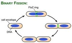
Binary fission is the form of asexual reproduction and cell
division used by all bacteria, some protozoa. This process results in the reproduction of prokaryotic cell by division into two parts similar to the original cell.
Binary fission accompanied with DNA replication.
division used by all bacteria, some protozoa. This process results in the reproduction of prokaryotic cell by division into two parts similar to the original cell.
Binary fission accompanied with DNA replication.
a) Budding
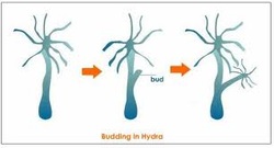
Budding is a form of asexual reproduction in which a new
organism grow on the original one. It stays attached while it grows. When it is fully grown , it detach from the original one.
organism grow on the original one. It stays attached while it grows. When it is fully grown , it detach from the original one.
a) Vegetative reproduction
Vegetative reproduction is a form of asexual reproduction in plants. It is a process by which new individuals arise without production of seeds.
a) Parthenogenesis (virgin creation)
It is a form of asexual reproduction in females, where growth and development of embryos occurs without fertlization by male. In plants, parthenogenesis means development of an embryo from an unfertilized egg cell. The offspring produced by parthenogenesis are always female in species.
Also, parthenogenesis occurs naturally in some invertebrate animal species as in water fleas, nematodes, some bees and some scorpion species. In vertebrate as some reptiles, fish and rarely birds and sharks this type of reproduction has been induced artificially. The parenthogenesis term is sometimes used inaccurately to describe reproduction modes in hermaphroditic species which can reproduce organs of both sexes.
Also, parthenogenesis occurs naturally in some invertebrate animal species as in water fleas, nematodes, some bees and some scorpion species. In vertebrate as some reptiles, fish and rarely birds and sharks this type of reproduction has been induced artificially. The parenthogenesis term is sometimes used inaccurately to describe reproduction modes in hermaphroditic species which can reproduce organs of both sexes.
e) Fragmentation
Fragmentation is a form of asexual reproduction where an organism is split into fragments. Each of these fragments develop into mature individuals that are similar to the original organisms. Fragmentation is caused by mitosis. Fragmentation is seen in molds, hydra, planaria and sea stars and many algae.
f) Sporogenesis
Reproduction via spores involves the spreading of the spores by water or air.Algae and some fungi use motile zoospores that swim to new locations before developing into sessile organisms.
Animal and reproduction
The word "animal" comes from anima, meaning soul ( the latin origin ). Usually, animal word refers to non-human animals. Frequently, it is closest to human such as vertebrates or mammals use. The biological definition of word refers to all members of the animal kingdom ranging from unicellular amoeba cell to human. Most animals are motile, meaning they can move spontaneously and independently.
Nearly all animals undergoe some form of sexual reproduction. Animals have a few specialized reproductive cells, motile spermatozoa or non-motile ova. These fuse to form fertilized cells (zygotes) which develop into new individuals.
Some animals are also capable of asexual reproduction. This may take place through parthenogenesis,where fertile eggs are produced without mating or in some cases through fragmentation.
Animals can be grouped into those which give birth to living offspring (viviparous) and those which lay eggs that eventually hatch into offspring (oviparous). The difference is in the place where the offspring develops before it is born.
The following table shows which groups are viviparous and which groups are oviparous.
Table: The vertebrates classified into viviparous and oviparous
groups.
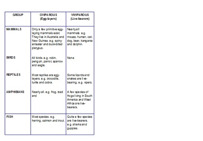
Amongst the invertebrates there are many which are oviparous but a few are viviparous such as sea anemones and aphids.
Sex Cells
A species of animal usually exists in two types or sexes called males and females. Each sex has its role to play in reproduction. When an animal gives birth or lays eggs, it is always the female which does this. In some cases, such as the sea horse and the midwife toad, the male appears to give birth to the offspring. In these exceptions the male has been given the eggs to look after but it is still the female animal that lays the eggs in the first place.
Sperm Cell
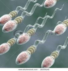
Sperm cells are very small but they are very specialized too.
The sperm cells of different species of animal are all about the same size, about 60µm long. All sperms have a head and a tail called a flagellum. They use the flagellum to swim through liquids. The head of the sperm is very important because it contains the nucleus. The nucleus carries half of the information needed for reproduction.
Sperm cells are made in very large numbers by special organs in the male's body called testes. In most animals the testes are carried inside the male's abdomen but in mammals they are kept in a sack of skin called the scrotum outside the abdomen.
Egg Cell
Egg cells are produced by female animals in special organs called ovaries. These are found inside the abdomen of all female animals.
Egg cells are much bigger and simpler than sperm cells in their structure. They do, however, vary a lot from one species of animal to another.
For example the egg of a human is only 0,1 mm wide but the egg of a chicken is 20 mm wide. Even so the human egg cell is still nearly 50 times wider than a
sperm cell. The reason for the difference in size is that the egg has a large food supply stored inside it called yolk. Yolk is the yellow part of a chicken's egg. The egg also has its own nucleus which carries the other half of the
information needed for reproduction.
Because eggs are bigger than sperms they are not produced in such large numbers. Even so animals which do not take care of their offspring, such as fishes and frogs, will lay a large number of eggs. Animals which take care of their offspring, such as mammals and birds, will produce less eggs.
Table: The number of eggs produced by each species
Species no. of egg produced at one time
Cod 5000000
Frog 1500
Viper 12
Human 1
_____________________________________________________
Reproduction In Fish
Three more important terms in the science of fish reproductive biology are Oviparous, Ovoviviparous and Viviparous.
Oviparous
if the eggs are fertilized internally and then laid by the female.
Ovoviviparous
if the eggs are fertilized internally and then carried in the females body until they hatch, then they are born alive, not laid as an egg. Still all the nutrients the young embyro needs are in the egg before it is fertilized.
Viviparous
if the eggs are fertilized internally, and the embryos kept within the females body until they are born alive, but these embryos receive nutrients directly from the mother while they are developing, in addition to those in the egg at the time of fertilization.
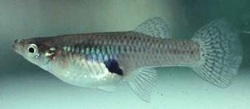
Internal fertilization is possible for fish via modification of the anal fin of the male into a copulatory organ. Viviparity is rare in fish (but common amongst mammals), a very successful example is the common Mosquito fish, Gambusia affinis, which produces about 30 young with a gestation period of 24 days.
In fish, the reproductive process involves three basic steps:
1- maturation (the development of the gametes to a point where fertilization can occur).
2- ovulation (the release of eggs from the ovary.
3- spawning (the diposition of eggs and sperm so that they can unite).
In fish, as in higher animals, hormones play a critical role in the reproductive process such as the hormones of pituitary gland. The primary tissues involved in this hormonal cascade are the hypothalamus, pituitary gland, and gonads.
Fish have evolved to reproduce under environmental conditions that are favorable to the survival of the young. Before spawning, seasonal cues begin the process of maturation. In many fish, this can take up to a year. When the gametes have matured, an environmental stimulus may signal the arrival of optimal conditions for the fry, triggering ovulation and spawning. Examples of environmental stimuli are changes in photoperiod,
temperature, rainfall, and food availability. A variety of sensory receptors detect these cues, including the eye, pineal gland, olfactory organs, taste buds, and thermoreceptors.
Furthermore, the hypothalamus is sensitive to signals from sensory receptors and releases hormones in response to environmental cues. Principal among these hormones are gonadotropin releasing hormones (GnRH), which travel from the hypothalamus to
the pituitary gland. Certain cells of the pituitary receive GnRH and release gonadotropic hormones into the bloodstream. The gonadotropic hormones travel to the gonads, which synthesize steroids responsible for final maturation of the gametes.
Maturation of the egg is attained by vitellogenesis process, in which yolk proteins are produced in the liver, transported to the ovary, and stored in the egg, resulting in tremendous egg enlargement. The yolk is important as a source of nutrition for the developing embryo.
The maturity of eggs can be determined using biopsy techniques. Eggs are removed
from the ovaries, cleared with a prepared solution, and viewed under a microscope. In mature eggs, the migration of the germinal vesicle to the side of the cell will be complete.
After the egg has matured, a class of compounds called prostaglandins are synthesized.
These stimulate ovulation, which is the rupture of the follicle cells that hold the egg. The egg is then released into the body cavity or ovarian lumen, where it may subsequently be released to the outside environment.
Hermaphroditic Fishes
Fishes producing both sperm and eggs, usually at different stages of its life are called self-fertilization fishes. however, they are probably rare.
In birds and mammals, hermaphroditism is usually a pathological condition causing
infertility. The most common vertebrate hermaphrodites are fishes, which
display several types of hermaphroditism.
Hermaphroditic fish species can be divided into three groups. The first are the synchronous hermaphrodites, in which ovaries and testicular tissues exist at the same time and in which both sperm and eggs are produced. One such species is Servanus
scriba.
In nature and in aquaria, these fish form spawning pairs. As soon as one of the fish spawns its eggs, the other fish fertilizes them. Then the fish reverse their roles, and the
fish that was formerly male spawns its eggs so that they can be fertilized by the sperm of its partner.
Oviparous
if the eggs are fertilized internally and then laid by the female.
Ovoviviparous
if the eggs are fertilized internally and then carried in the females body until they hatch, then they are born alive, not laid as an egg. Still all the nutrients the young embyro needs are in the egg before it is fertilized.
Viviparous
if the eggs are fertilized internally, and the embryos kept within the females body until they are born alive, but these embryos receive nutrients directly from the mother while they are developing, in addition to those in the egg at the time of fertilization.
Internal fertilization is possible for fish via modification of the anal fin of the male into a copulatory organ. Viviparity is rare in fish (but common amongst mammals), a very successful example is the common Mosquito fish, Gambusia affinis, which produces about 30 young with a gestation period of 24 days.
In fish, the reproductive process involves three basic steps:
1- maturation (the development of the gametes to a point where fertilization can occur).
2- ovulation (the release of eggs from the ovary.
3- spawning (the diposition of eggs and sperm so that they can unite).
In fish, as in higher animals, hormones play a critical role in the reproductive process such as the hormones of pituitary gland. The primary tissues involved in this hormonal cascade are the hypothalamus, pituitary gland, and gonads.
Fish have evolved to reproduce under environmental conditions that are favorable to the survival of the young. Before spawning, seasonal cues begin the process of maturation. In many fish, this can take up to a year. When the gametes have matured, an environmental stimulus may signal the arrival of optimal conditions for the fry, triggering ovulation and spawning. Examples of environmental stimuli are changes in photoperiod,
temperature, rainfall, and food availability. A variety of sensory receptors detect these cues, including the eye, pineal gland, olfactory organs, taste buds, and thermoreceptors.
Furthermore, the hypothalamus is sensitive to signals from sensory receptors and releases hormones in response to environmental cues. Principal among these hormones are gonadotropin releasing hormones (GnRH), which travel from the hypothalamus to
the pituitary gland. Certain cells of the pituitary receive GnRH and release gonadotropic hormones into the bloodstream. The gonadotropic hormones travel to the gonads, which synthesize steroids responsible for final maturation of the gametes.
Maturation of the egg is attained by vitellogenesis process, in which yolk proteins are produced in the liver, transported to the ovary, and stored in the egg, resulting in tremendous egg enlargement. The yolk is important as a source of nutrition for the developing embryo.
The maturity of eggs can be determined using biopsy techniques. Eggs are removed
from the ovaries, cleared with a prepared solution, and viewed under a microscope. In mature eggs, the migration of the germinal vesicle to the side of the cell will be complete.
After the egg has matured, a class of compounds called prostaglandins are synthesized.
These stimulate ovulation, which is the rupture of the follicle cells that hold the egg. The egg is then released into the body cavity or ovarian lumen, where it may subsequently be released to the outside environment.
Hermaphroditic Fishes
Fishes producing both sperm and eggs, usually at different stages of its life are called self-fertilization fishes. however, they are probably rare.
In birds and mammals, hermaphroditism is usually a pathological condition causing
infertility. The most common vertebrate hermaphrodites are fishes, which
display several types of hermaphroditism.
Hermaphroditic fish species can be divided into three groups. The first are the synchronous hermaphrodites, in which ovaries and testicular tissues exist at the same time and in which both sperm and eggs are produced. One such species is Servanus
scriba.
In nature and in aquaria, these fish form spawning pairs. As soon as one of the fish spawns its eggs, the other fish fertilizes them. Then the fish reverse their roles, and the
fish that was formerly male spawns its eggs so that they can be fertilized by the sperm of its partner.
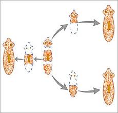
In the following figure the gonadal changes in the hermaphroditic fish Sparus auratus, (A) the male phase, (B) the transitory phase, and (C) the final, female phase.
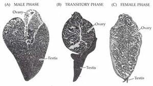
In other hermaphroditic species, an individual undergoes a genetically programmed sex change during its development. In these cases, the gonads are dimorphic, having both male and female areas. One or the other is predominant during a certain phase of life. In protogynous ("female-first") hermaphrodites, an animal begins its life as a female, but later becomes male. The reverse is the case in protandrous ("male-first") species.
Inducing Reproduction in Fish
There are two main strategies used to induce reproduction. The first is to provide an
environment similar to that in which spawning occurs naturally. Catfish, for example, like to spawn in enclosed spaces such as hollow logs. A farmer can simulate this by putting milk cans in a pond. The presence of vegetation and an increase in temperature will usually work for goldfish. Changing the photoperiod in a hatchery can accelerate or delay maturation and ovulation in many salmon and trout species.
The second strategy is to inject the fish with one or more naturally occurring reproductive hormones or their synthetic analogs. This is only effective in fish that are already in breeding condition and have mature eggs in which the germinal vesicle has migrated.
Often the two strategies are used sequentially; the first to manipulate maturation,
then the second to induce ovulation.
Methods of inducing spawning in fishes
Inducing spawning differ from species to species. They will also differ according to the
goals and means of the farmer. It must be emphasized that the following techniques do not apply to all situations.
Injecting the Fish
There are two common places to inject hormones into a fish.
An intraperitoneal injection (i.p.)
An intraperitoneal (within the body cavity) injection is given through the ventral (bottom) part of the fish behind either the pelvic or pectoral fin.
Intramuscular injection (i.m.)
Intramuscular (within the muscle) injections are commonly done on the dorsal (upper) part of the fish above the lateral line and below the anterior part of the dorsal fin.
In either case, it is important to place the needle so that it slides under the scale rather than through it.
Two dosage levels are commonly used (a preparatory dose and a decisive, or final, dose with a time gap generally of 12 to 24 hours between the two injections).
The preparatory dose brings the fish to the brink of spawning, and the decisive dose
induces ovulation. In general, the preparatory dose is about 10 percent of the total dose. For some fish, several preparatory doses may be necessary.
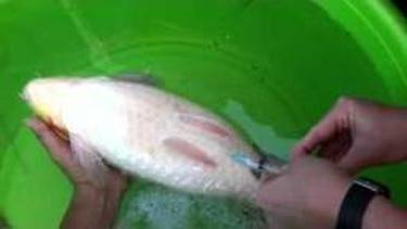
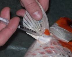
Stripping of Fishes
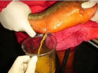
The female should be held around the caudal fin with one hand, while applying slight pressure to the abdomen with the other hand. If ovulation has occurred, a stream of eggs will emerge. If only a few appear, the female is still “green” and should be returned to the holding tank. If there is a stream of eggs, the abdomen should be messaged from front to back to strip out all the eggs.
Reproduction in amphibians
Reproduction of frogs
Once adult frogs reach maturity, they will assemble at a water source to breed. Many frogs return to the bodies of water where they were born, often resulting in annual migrations involving thousands of frogs. Once at the breeding ground, male frogs call to attract a mate, collectively becoming a chorus of frogs. The call is unique to the species, and will attract females of that species. Some species have satellite males who do not call, but intercept females that are approaching a calling male.
The male and female frogs then undergo amplexus. This involves the male mounting the female and gripping her (sometimes with special nuptial pads) tightly. Fertilization external: the egg and sperm meet outside of the body. The female releases her eggs, which the male frog covers with a sperm solution. The eggs then swell and develop a protective coating. The eggs are typically brown or black, with a clear, gelatin-like covering.
Frog Spawning
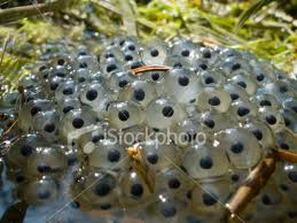
Most temperate species of frogs reproduce between late autumn and early spring. Reproducing in these conditions helps the developing tadpoles because dissolved oxygen concentrations in the water are highest at cold temperatures. More importantly, reproducing early in the season ensures that appropriate food is available to the developing frogs at the right time.
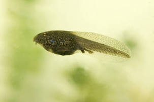
Eggs hatch and continue life as tadpoles
Some species of frog lay eggs on the forest floor and protect them, guarding the eggs from predation and keeping them moist. The frog will urinate on them if they become too dry. After hatching, a parent (the sex depends upon the species) will move them, on its back, to a water-holding flowering plant. The parent then feeds them by laying unfertilized eggs in the bromeliad until the young have metamorphosed. Other frogs carry the eggs and tadpoles on their hind legs or back. Some frogs even protect their offspring inside their own bodies.
Reproduction In Reptile
Mating rituals in reptiles
Many reptiles display mating rituals as change of their colours as lizards. Male turtles have been know to woo a female by bobing his head or using his claws to caress her face.
Patterns of reproduction in reptiles
Most reptiles reproduce sexually, though some are of asexual reproduction.
Sexual reproduction in reptiles
All reproduction is carried out through the cloaca. Cloaca is the single entrance at the base of tail , also, by which waste is eliminated. Most reptile have copulatory organs and some lack copulatory organ.
In turtles and crocodilians, the male has a single median penis, while snaks and lizards possess a pair of hemipenes.
However, tuataras lack copulatory organs, so the male and female press their cloacas
together (cloacal kiss) as the male to excretes sperms.
In reptiles, there is an additional canal between the testis and the head of the epididymis, which receives the various efferent ducts. This is, however, absent in all birds and mammals.
The oviducts of some female reptiles are capable of storing sperm in viable condition for months or even years. In some turtles and snakes, fertilization can occur three years after insemination.
Most reptiles lay amniotic eggs covered with leathery or calcareous shells. An amnion, chorion and allantois are present during embryonic life. There are no larval stages of development.
Fig.: Amniotic eggs of reptiles with leathery shells.
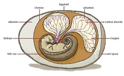
Some species retain the eggs until just before hatching, others provide maternal nourishment to supplement the yolk, and yet others lack any yolk and provide all nutrients via a structure similar to mammalian placenta.
Asexual reproduction in reptiles
Asexual reproduction has been identified in squamates in 6 families of lizards and one family of snake. In which a population of females is able to produce a unisexual diploid clone of the mother.
This is not a widespread mode of reproduction in reptiles, but it is known to occur in several species of lizards and at least one snake.
Facultative parthenogenesis has only recently been discovered among captive reptiles, and there is as yet no information on whether it occurs in nature.
Egg hatching
The number of eggs yeilded during reproduction varies depending upon the type of reptile. Reptiles can lay as few as one egg or over hundred according to its type. African tortoises can lay 1-2 eggs at a time, while some sea turtles lay up to 150 eggs at once in nests. The reptiles then leave the eggs to hatch on their own from sand, leaves or soil.
Reproduction in mouse
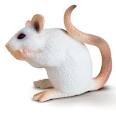
Mice are common experimental animals in biology and psychology; primarly because they are mammals, easy to maintain and handle, reproduce quickly. In addition to being small, relatively inexpensive and easily maintained, there are further benefits to the use of mice in laboratory research. Because mice can reproduce quickly, several generations of mice can observed in a relatively short period of time.
Most laboratory mice are hybrids of different subspecies, most commonly of Mus musculus domesticusand Mus musculus musculus.
Laboratory mice come in a variety of coat colours including agouti, black and albino. Many laboratory
strains are inbred so as to make them genetically almost identical. The different strains are identical with specific letter-digit combinations for example C57BL/6 and BALB/c.
Female mice have an estrous cycle that is 4-6 days long. If several females are maintained under crowded conditions, they will often not have an estrous at all. If they are then exposed to male urine, they will become estrous after 72 hours.
Male mice court females by emitting characteristic ultrasonic calls in the 30kHz-110kHz range. The calls are most frequent during courtship when the male is sniffing and following the females; however , the calls continue after mating has begun at which time the calls are concident with mounting behaviour. Males can induced to emit these calls by female pheromones.
The vocalizations appear to be different individuals and have been compared to bird songs becauase of their complexity. While females have the capability to reproduce ultrasonic calls, they typically do not do so during mating behaviour.
Fig. : A baby mouse, 4 days old.
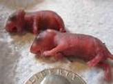
Following copulation, female mice will normally develop a vaginal plug which prevents further copulation.
This plug stays in place for some 24 hours. The gestation period is about 19-21 days, and they give birth to a litter of 3-14 young (avarage 6-8). One female can have some 5-10 litters per year, so their population can increase very quickly. Breeding occurs throughout the year (however, animals living in the wild don't reproduce in the colder months, even though they don't hibernate). The newborn are blind and witout fur.
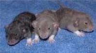
Fur starts to grow some three days after birth and the eyes open one to two weeks after birth. Females reach sexually maturity at about 6 weeks and males at about 8 weeks, but both
can breed as early as five weeks.
This plug stays in place for some 24 hours. The gestation period is about 19-21 days, and they give birth to a litter of 3-14 young (avarage 6-8). One female can have some 5-10 litters per year, so their population can increase very quickly. Breeding occurs throughout the year (however, animals living in the wild don't reproduce in the colder months, even though they don't hibernate). The newborn are blind and witout fur.
Fur starts to grow some three days after birth and the eyes open one to two weeks after birth. Females reach sexually maturity at about 6 weeks and males at about 8 weeks, but both
can breed as early as five weeks.
Fig.: 2 weeks old, just about to open its eyes.
House mice usually live under a year in the wild. This is due to a high level of predation and exposure to harsh environments. In protected environments, however , they often live two to three years.
Reproduction in Human
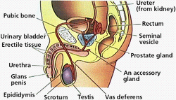
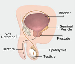
(I) Male Reproductive System
Its functions are:
-Production of sperms
-Sperm delivary to the female reproductive tract.
-Production of Hormones.
Components of Male Reproductive System
The male reproductive system consists of:
1- A pair of testes that produce sperm.
2- Ducts that transport the sperm to the penis.
--3- Glands that add secretions to the sperm to make semen.
Testis
- Each testis contains about 20 meters of seminiferous tubules, where spermatogenesis occurs.
- Sperm need temperatures between 2 to 10 oC lower than the body temperature to develop.
- This is the reason where the testes are located in the scrotal sacs (or scrotum) that hangs outside the body to further reduce its temperature.
--- The sperm are formed by meiosis in the seminiferous tubules.
- When the sperm are mature they accumulate in the collecting ducts and then pass to the epididymis before moving to the vas deferens. The two sperm ducts join the urethra.
- Ejaculation discharges the semen from the urethera by the contraction of the epididymis, vas deferens, prostate gland and urethra.
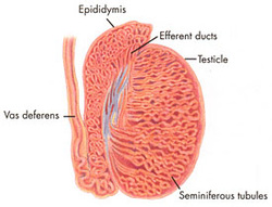
Epididymis
The epididymis is part of the male reproductive system and is present in all male amniotes. It is a narrow, tightly-coiled tube connecting the efferent ducts from the rear of each testicle to its vas deferens. A similar, but probably non-homologous, structure is found in cartilaginous fishes.
The epididymis can be divided into three main regions:
- The head (caput). The head of epididymis recieves spermatozoa via efferent ducts of the mediastinum of the testis. It is characterized histologically by a thin myoepithilium. the concentration of the sperm here is dilute.
- the body (Corpus).
- The tail (Cauda). This has a thicker myoepithelium than the head region, as it is involved in absorbing fluid to make the sperm more concentrated.
Role in storage of sperm and ejaculant
Spermatozoa formed in the testis enter the caput epididymis, progress to the corpus, and finally reach the cauda region, where they are stored. Sperm entering the caput epididymis are incomplete - they lack the ability to swim forward (motility) and to fertilize an egg. During their transit in the epididymis, sperm undergo maturation processes necessary for them to acquire these functions.
Final maturation is completed in the female reproductive tract (capacitation).
During ejaculation, perm flow from the lower portion of the epididymis (which functions as a storage reservoir). They have not been activated by products from the prostate gland, and they are unable to swim, but are transported via the peristaltic action of muscle layers within the vas deferens, and are mixed with the diluting fluids of the seminal vesicles and other accessory glands prior to ejaculation (forming semen).
Its functions are:
-Production of sperms
-Sperm delivary to the female reproductive tract.
-Production of Hormones.
Components of Male Reproductive System
The male reproductive system consists of:
1- A pair of testes that produce sperm.
2- Ducts that transport the sperm to the penis.
--3- Glands that add secretions to the sperm to make semen.
Testis
- Each testis contains about 20 meters of seminiferous tubules, where spermatogenesis occurs.
- Sperm need temperatures between 2 to 10 oC lower than the body temperature to develop.
- This is the reason where the testes are located in the scrotal sacs (or scrotum) that hangs outside the body to further reduce its temperature.
--- The sperm are formed by meiosis in the seminiferous tubules.
- When the sperm are mature they accumulate in the collecting ducts and then pass to the epididymis before moving to the vas deferens. The two sperm ducts join the urethra.
- Ejaculation discharges the semen from the urethera by the contraction of the epididymis, vas deferens, prostate gland and urethra.
Epididymis
The epididymis is part of the male reproductive system and is present in all male amniotes. It is a narrow, tightly-coiled tube connecting the efferent ducts from the rear of each testicle to its vas deferens. A similar, but probably non-homologous, structure is found in cartilaginous fishes.
The epididymis can be divided into three main regions:
- The head (caput). The head of epididymis recieves spermatozoa via efferent ducts of the mediastinum of the testis. It is characterized histologically by a thin myoepithilium. the concentration of the sperm here is dilute.
- the body (Corpus).
- The tail (Cauda). This has a thicker myoepithelium than the head region, as it is involved in absorbing fluid to make the sperm more concentrated.
Role in storage of sperm and ejaculant
Spermatozoa formed in the testis enter the caput epididymis, progress to the corpus, and finally reach the cauda region, where they are stored. Sperm entering the caput epididymis are incomplete - they lack the ability to swim forward (motility) and to fertilize an egg. During their transit in the epididymis, sperm undergo maturation processes necessary for them to acquire these functions.
Final maturation is completed in the female reproductive tract (capacitation).
During ejaculation, perm flow from the lower portion of the epididymis (which functions as a storage reservoir). They have not been activated by products from the prostate gland, and they are unable to swim, but are transported via the peristaltic action of muscle layers within the vas deferens, and are mixed with the diluting fluids of the seminal vesicles and other accessory glands prior to ejaculation (forming semen).
Fig.: Showing epididymis,efferent ducts, vas deferens, seminifereous tubule.
The epididymis possesses numerous, long atypical microvilli. These processes are often called stereocillia; this is incorrect, as they neither contain the microtubular structures of cilia nor function like cilia.
In pathology: An inflammation of the epididymis is called epididymitis.
As a vestigial structures: A Gartner's duct is a homologous remnant in the female.
In the embryo, the epididymis develops from tissue that once
formed the mesonephros, a primitive kidney found in many aquatic vertebrates. Persistence of the cranial end of the mesonephric duct will leave behind a remnant called the appendix of the epididymis. In addition, some mesonephric tubules can
persist as the paradidymis, a small body caudal to the efferent ductules.
Epididymectomy: This is the surgical removal of the Epididymis carried out under local anaesthesia. This is most often performed to relieve pain associated post-Vasectomy.
Seminal vesicle
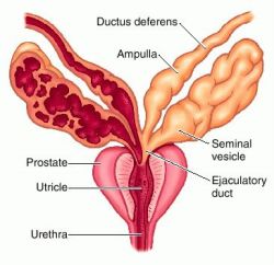
Either of a pair of pouchlike glands situated on each side of the male urinary bladder that secrete seminal fluid and nourish spermatozoa. Seminal fluid promote the movement of spermazoa through the urethera.
Anatomy: Each seminal gland spreads approximately 5 cm,though the full length of seminal vesicle is approximately 10 cm, but it is curled up inside of the gland's structure. Each gland forms as an outpocketing of the wall of ampulla of each vas deferens.Theexcretory duct of seminal gland opens into the vas deferens as it enters the prostate gland.
Function: The seminal vesicles secrete a significant proportion of the fluid that
ultimately becomes semen. Lipofuscin granules from dead epithelial cells gives the secretion its yellowish color. About 60% of the seminal fluid in humans originates from the seminal vesicles, but is not expelled in the first ejaculatefractions which are dominated by spermatozoa and zinc-rich prostatic fluid. The excretory duct of each seminal gland opens into the corresponding vas deferens as it enters the prostate gland. Seminal vesicle fluid is alkaline along with the prostatic fluid, resulting in human semen having a mildly alkaline pH. The alkalinity of semen helps neutralize the acidity of the vaginal tract, prolonging the lifespan of sperm. Acidic ejaculate (pH <7.2) may be associated with blockage of seminal
vesicles. The thick secretions from the seminal vesicles contain protein, enzymes,fructose,mucus,vitamin C,flavins,phosphorylcholineand prostaglandins. The high fructose concentrations provide nutrient energy for the spermatozoa when stored in semen in the laboratory. Spermatozoa ejaculated in the vagina are not likely to have contact with the seminal vesicular fluid but transfer directly from the prostatic fluid
into the cervical mucus as the first step on their travel through the female reproductive system. The fluid is expelled under sympathetic contraction of the muscularis muscle coat. In vitro studies have shown that sperm expelled together with seminal vesicular fluid show poor motility and survival, and the sperm chromatin is less protected. Therefore the exact physiological importance of seminal vesicular fluid is not clear. It has been speculated that it is a developmental rest, still seen among some rodents where the last part of the ejaculate form a spermicidal plug which reduces the chances for sperm from a later arriving male to proceed to the oocyte.
prostate
The prostate gland surrounds the urinary passage at the exit of the male bladder.
The gland is very small in babies and grows at the time of puberty in response
to testosterone secreted by the testicles. The function of the prostate is to secrete fluid which together with secretions from the seminal vesicles makes up most of the volume of the seminal fluid. The functions of seminal fluid are incompletely
understood and more than a hundred compounds have been isolated from it. The
gland has given its name to the group of substances known as prostaglandins,
first identified at this site, but now known to be present throughout the body;
prostaglandins in the seminal fluid may cause contractions of the female genital tract, facilitating transport of sperm through the uterus to the Fallopian tubes. The fluid also helps with the nutrition of sperm and defence against infection. The ejaculate forms a clot which sticks to the mucus of the cervix, enabling the passage of sperm into the mucus, through which they can travel to enter the uterus. An enzyme called prostatic specific antigen (PSA) then liquefies the sperm
clot and the seminal fluid subsequently seeps out of the vagina. PSA is produced
by the lining cells of prostate ducts and a small amount can be detected
circulating in the blood. Any condition that increases prostate cells, such as
benign enlargement of the prostate, or cancer, or any condition which causes
leakiness of the cells such as prostatitis, results in increased levels of PSA
in the blood. Very high levels of PSA usually indicate cancer, but moderately
raised levels may indicate a whole variety of prostate disorders. There is
worldwide research on the merits of PSA estimation as a screening test for
prostate cancer but because increased levels may occur in a number of prostate
disorders it can never be a perfect test.
Most men remain unaware of their prostate until late middle age when enlargement interferes with urination by constricting the urethra and reducing urine flow. This is often associated with the need to rise at night and pass urine more frequently. For more minor symptoms, there is effective drug treatment. When the symptoms become sufficiently disabling the usual treatment is an operation. The current lifetime chance of requiring a prostate operation is about one in ten. Prostate cancer is very common in elderly men and is a significant cause of premature death, but the paradox is that many more men have prostate cancer than die of it. Benign enlargement of the prostate and in some cases, cancer, can be treated by
operations to remove part, or in cancer cases, the whole of the prostate. Many
men fear these operations will result in impotence.
In most cases of benign enlargement of the prostate there is no danger of
impotence. Removal of part or all of the prostate does however result in a lack
of external ejaculation because the junctions between the genital and urinary
tracts lie within the prostate. In operations to remove the gland completely, it
is possible for the nerves to the penis to be damaged, since they run close to
the prostate. For many men with prostate cancer, total removal by operation is
not feasible and the mainstay of treatment is to give hormones. Both the normal
and the cancerous prostate grow in response to testosterone and the strategy of
hormone treatments is to deprive the cancer of male hormone. This can be done in
a number of different ways, both by blocking production of testosterone and by
blocking its action. Hormone treatments are not curative but they may remain effective for many years.
Bulbourethral gland (Cowper's gland)
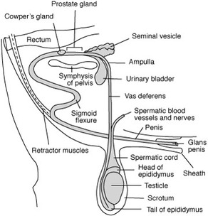
It is also, called a Cowper's gland (according to anatomist William Cowper). It is one of two small exocrine gland present in human male.
Either of two small racemose glands that are located below the prostate and discharge a component of the seminal fluid into the urethra. They are homologous to the Bartholin's glands in the female.
Location:
Bulbourethral glands are located posterior and lateral to the membranous portion of the urethra at the base of the penis, between the two layers of the fascia of the
urogenital diaphragm, in the deep perineal pouch. They are enclosed by transverse fibers of the sphincter urethrae membranaceae muscle.
Function:During sexual arousal each gland produces a clear, viscous secretion known as pre-ejaculate. This fluid helps to lubricate the urethra for spermatozoa to pass through, neutralizing traces of acidic urine in urethra, and helps flush out any residual urine or foreign matter. It is possible for this fluid to pick up sperm, remaining in the urethral bulb from previous ejaculations, and carry them out prior to the next ejaculation. The Cowper's gland also produces some amount of prostate-specific antigen (PSA), and Cowper's tumors may increase PSA to a level that makes prostate cancer suspicious.
vas deferens
It is also, called ductus deferens ( carrying away vessel by Latin meaning). It is part of the male anatomy of many vertebrates; they transport sperm from the epididymis to
the ejaculatory duct.
The tube on each side which leads from the testisto the urethra, carrying sperm and some other
components of seminal fluid. Each vas ends by joining the duct of a seminal vesicle, to enter the urethra (the urinary passage) where that traverses the prostate gland. May be closed off by the operation of vasectomy, preventing sperm from entering the ejaculate.
Function: During ejaculation the smooth muscle in the walls of the vas deferens contracts
reflexively, thus propelling the sperm forward. This is also known as peristalsis. The sperm is transferred from the vas deferens into the urethra, collecting secretions from the male accessory sex glands such as the seminal vesicles, prostate gland and the bulbourethral glands, which form the bulk of semen.
Vas deferens and contraception :
The procedure of deferentectomy, also known as a vasectomy, is a method of contraception in which the vasa deferentia are permanently cut, though in some cases it can be reversed. A modern variation, which is also known as a vasectomy even though it does not include cutting the vas, involves injecting an obstructive material into the ductus to block the flow of sperm. In either procedure, active sperm may be still be present in the seminal vesicles for as long as 12 weeks.
Blood supply of vas deferens:
The vas deferens is supplied by an accompanying artery (artery of vas deferens). This artery normally arises from the inferior vesical artery, a branch of the internal iliac artery.
Variation among vertebrates
In cartiliginous fishes and amphibians, sperm is carried through the archinephric duct, which also partially helps to transport urine from the kidneys.
In teleosts, there is a distinct sperm duct, separate from the ureters, and often called the vas deferens, although probably not truly homologous with that in humans.
In amniotes, however, the chinephric duct has become a true vas deferens, and is used only for conducting sperm, never urine. As in cartilaginous fish, the upper part of the duct forms the epididymis. In many species, the vas deferens ends in a small sac for storing sperm.
Primitive jawless fishes, the only vertebrates to lack any structure resembling a vas deferens which release sperm directly into the body cavity, and then into the surrounding water through a simple opening in the body wall.
Pathology: The vas deferens may be obstructed, or may be completely absent
(cystic fibrosis), causing male infertility.
Spermatogenesis (Production Of Sperms)
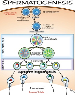
The walls of the seminiferous tubules consist of diploid spermatogonia, stem cells that are the precursors of sperm.
-Spermatogonia divide by mitosis to produce more differentiated spermatocytes.
- Each spermatocyte produces by meiosis 4 haploid spermatids. This process takes about three weeks to complete.
- Then the spermatids differentiate into sperm, losing most of their cytoplasm.
Sertoli cell
The Sertoli cell is the 'nurse' cell of the testes. Its main function is concerned with nurturing the developing sperm cells during the process of spermatogenesis.
Sertoli cells secrete anti-Mullerian hormone (AMH) during the early stages of fetal life.
After puberty, they secrete the hormone inhibin.
The junctions of Sertoli cells form the blood-testis barrier, a structure that partitions the interstitial blood compartment of the testis from the adluminal compartment of the seminiferous tubules. Sertoli cells control the entry and exit of nutrients,hormones and other chemicals into the
tubules of the testis. Once fully differentiated the Sertoli cell is unable to proliferate and therefore
once spermatogenesis has begun no more Sertoli cells are created. Recently however, some scientists have found a way to grow these cells outside of the body. This gives rise to the possibility of repairing some defects that cause male infertility.
Sperm
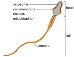
Sperm Structure: Sperm cell
consists of :
- A head, which has an acrosome at its tip and contains
a haploid set of chromosomes. An acrosome at the head tip produces enzymes that help penetrate the female ovum (egg).
- A midpiece containing mitochondria and a single centriole.
- A tail.
Quantity Of Sperm/Day
- An adult male produces about 150 million sperm cells /day.
- These move into the epididymis.
- In epididymis they undergo further maturation in acidic environment and keeps the mature sperm inactive.
Hormones Of Male Reproductive System
1- Testosterone.
2- Interstitial Cell Stimulating Hormone (ICSH)
3- Leutinizing Hormone (LH).
4- Folliclle Stimulating Hormone (FSH).
Testosterone
- Testosterone is made in the interstitial cells.
- The interstitial cells lie between the seminiferous tubules.
Interstesial Cell Stimulating Hormone (ICSH)
Interstitial cells are the targets for LH hormone. It is identical to LH found in female.
Leutinizing Hormone (LH)
LH is a product of the anterior lobe of the pituitary gland.
Folliclle Stimulating Hormone (FSH)
FSH acts directly on spermatogonia to stimulate sperm production.
Also, it is produced by anterior lobe of pituitary gland.
Parasympathetic Nerves and Penile Erection
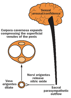
Psychogenic stimuli from the brain or from tactile receptors on the penis (via the pudendal nerve) activate efferent parasympathetic fibers of the pelvic nerve.
This stimulates vasodilatation increasing blood flow into the corpora cavernosa and to a lesser extent in the corpus spongiosum. This involves the neurotransmitters, acetylcholine and vasoactive polypeptide and increased nitric oxide production. The sympathetic outflow of the hypogastric nerve normally maintains myogenic tone in the flaccid penis but this is counteracted by the parasympathetic activity during sexual arousal. Prostaglandin E1 or a synthetic analog or α-adrenergic
blockers can reduce tone and cause tumescence.
