Comparative Circulation
Circulatory system and blood circulation is important for the survival in humans or animals. For proper strategies of blood circulation management we need information on nutrients and waste products delivery in any animal which depends on circulatory system or diffusion from unicellular animal to multicellular animals respectively.
Function of Circulatory System
The circulatory system performs many different functions:
1- It is responsible for carrying oxygen to cells of the body.
2- It also delivers nutrients to produce heat and energy.
3- It acts as a delivery system for hormones to specific target organs.
4- It performs by collecting metabolic waste and delivering it to excretory organs.
5- It plays a protective role by combating infections and helping to establish an immune system defense.
Thereare three types of circulatory systems (from simplest to most complex):
1.No circulatory System animals.
2.Open circulatory system animals.
3.Closed circulatory system animals.
Animals without circulatory system
These animals like the flatworms exhibit no circulatory system, but they are able to obtain nutrients, water, and oxygen by diffusion.
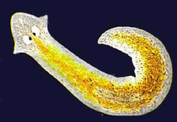
Fig.: Animals with No Circulatory System
Flat worms have no circulatory nor a respiratory system.
It is flat enough to allow these functions to occur at the cellular level and perform exchanges directly with its suroundings and gut.
Animals With Open Circulatory System
The open circulatory system can be found in arthropods and most mollusks and contains no capillaries or veins.
In it a heart pumps blood (hemolymph) through arteries and into spaces around the organs. This allows tissues to
exchange materials with the hemolymph, which is then drawn back into the heart as it relaxes.
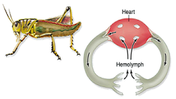
Fig.: Open Circulatory System
In open circulatory animals, hemolymph circulated by simple body movements, squeezes fluid through sinuses .
Hemolymph enters heart through ostia (pores). Relaxation of heart causes hemolymph to be drawn in; expelled as heart
contracts (creating vacuum).
Animals with closed circulatory system
In the closed type of circulatory system, the blood remains inside the blood vessels and does not come out. The blood flows from arteries to veins through small blood vessels called capillaries.
The closed type of circulatory system occurs in most of the Annelids, Cephalopods and Vertebrates including man.
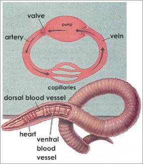
Fig.: Closed Circulatory System
The open and closed circulatory system in locust and earthworm respectively are indicated in the following Figure.
The open and closed circulatory system in locust and earthworm respectively are indicated in the following Figure.
Fig.: Open and Closed Circulatory Systems
The systems of fish, amphibians, reptiles, and birds show various stages of the evolution of the circulatory system.
In fish, the system has only one circuit, with the blood being pumped through the capillaries of the gills and on to the capillaries of the body tissues. This is known as single cycle circulation.
In reptiles, the ventricular septum of the heart is incomplete and the pulmonary artery is equipped with a sphincter muscle.
Birds and mammals show complete separation of the heart into two pumps, for a total of four heart chambers; it is thought that the four-chambered heart of birds evolved independently from that of mammals.
Human Blood Circulation
The human circulatory system is an organ system that permits blood and lymph circulation to transport nutrients (such as
amino acids and electrolytes), oxygen, carbon dioxide, hormones, blood cells, etc. to and from cells in the body to nourish it and help to fight diseases, stabilize body temperature and pH, and to maintain homeostasis.
Fig.: Human Circulatory System
The main components of the human cardiovascular system are the heart and the blood vessels.
It includes:
The pulmonary circulation, a "loop" through the lungs where blood is oxygenated; and the systemic
circulation, a "loop" through the rest of the body to provide oxygenated blood. An average adult contains five to six quarts (roughly 4.7 to 5.7 liters) of blood, which consists of plasma, red blood cells, white blood cells, and platelets.
Deoxygenated blood collects in two major veins: the superior vena cava and the inferior vena cava. The superior and inferior vena cava empty into the right atrium. The right atrium is the larger of the two atriums because it needs to be able to hold the larger amount of blood coming from the body.
The blood is then pumped through the tricuspid atrioventicular valve into the right ventricle.
From the right ventricle, blood is pumped through the pulmonary semi-lunar valve into the pulmonary trunk.
The deoxygenated blood leaves the heart by the pulmonary arteries and travels through the lungs (where it is oxygenated) and into the pulmonary vein. The oxygenated blood then enters the left atrium. The blood then travels through
the bicuspid valve, or mitral valve, into the left ventricle.
Elements Of Circulatory System
The heart is composed of cardiac muscle , an involuntary muscle (myocardium). The heart is exciting tissue with self surrounded by fibrous superficial and it deeply serous pericardium layers.It consists of four chambers right and left ventricles and right and left atria.
The main components of the human cardiovascular system are the heart and the blood vessels.
It includes:
The pulmonary circulation, a "loop" through the lungs where blood is oxygenated; and the systemic
circulation, a "loop" through the rest of the body to provide oxygenated blood. An average adult contains five to six quarts (roughly 4.7 to 5.7 liters) of blood, which consists of plasma, red blood cells, white blood cells, and platelets.
Deoxygenated blood collects in two major veins: the superior vena cava and the inferior vena cava. The superior and inferior vena cava empty into the right atrium. The right atrium is the larger of the two atriums because it needs to be able to hold the larger amount of blood coming from the body.
The blood is then pumped through the tricuspid atrioventicular valve into the right ventricle.
From the right ventricle, blood is pumped through the pulmonary semi-lunar valve into the pulmonary trunk.
The deoxygenated blood leaves the heart by the pulmonary arteries and travels through the lungs (where it is oxygenated) and into the pulmonary vein. The oxygenated blood then enters the left atrium. The blood then travels through
the bicuspid valve, or mitral valve, into the left ventricle.
Elements Of Circulatory System
- Heart
- Blood vessels
- Blood
The heart is composed of cardiac muscle , an involuntary muscle (myocardium). The heart is exciting tissue with self surrounded by fibrous superficial and it deeply serous pericardium layers.It consists of four chambers right and left ventricles and right and left atria.
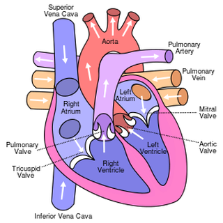
Fig.: Mammalian Heart
Right and Left Atria
The right atrium recieves deoxygenated blood from superior and inferior vena cava.
The left atrium recieing oxygenated blood from pulmonary veins.
Right and Left Ventricles
The right ventriclepumps deoxygenated blood into pulmonary circulation for the lungs.
The left ventricle pumps oxygenated blood into the systemic circulation for the body.
- Ventricles have thicker walls than the atria.
- The left ventricle have thicker walls because it needs blood pressure to pump blood to the whole body.
Cardiac Action Potential
Fig.: Cardiac Action Potential
If the membrane potential is at its baseline (about -85 mV), all Na+ channels are closed. A typical action potential is initiated when the membrane is depolarized sufficiently, and excitation will open Na+ channels, causing a large influx of Na+ ions.
The inward flow of Na+ ions increases the concentration of positively-charged cations in the cell and causes depolarization.
The efflux of K+ ions decreases the membrane potential or hyperpolarizes the cell.
Na+ inactivation is accompanied by opening of slowly Ca2+ channels at the same time as a few fast K+ channels open.
The balance between the outward flow of K+ and the inward flow of Ca2+ causes a plateau, its length (150 ms).
The delayed opening of additional Ca2+-activated K+ channels as the Ca2+ channels close, terminates the plateau and leads to repolarisation.
This "plateau" phase of the cardiac action potential is sustained by a balance between inward movement of Ca2+ through Ca++ channels and outward movement of K+ ions.
Role of Na+, K+ and Ca++ On Heart Rate
An excess of K+ ions in the extracellular environment markedly reduce the heart rate as well as the strength of contraction.
On the other hand, spastic contraction of the heart results from the presence of excess Ca++ ions.
Excessive levels of Na+ ions result in depression of cardiac function.
At the other extreme, a deficiency of Na+ ions in the extracellular environment leads to the development of a potentially
lethal condition called cardiac fibrillation.
Human cardiovascular system have double circulation:
- A pulmonary circuit to the lungs
- A systemic circuit to the body.
Fig.: Pulmonary and Systemic Circulation
Pulmonary Circulation
The pulmonary artery arises from the right ventricle and tranports deoxygenated blood (oxygen-poor) to the lungs, where the blood becomes oxygenated again. The four pulmonary veins return the oxygenated blood (oxygen-rich) to the left atrium of the heart. The pulmonary circulation is also referred to as the lesser circulation. The summary of the pulmonary circulation is thus:
Right Ventricle ------- pulmonary artery --------- lungs ------- pulmonary veins ------- left atrium --------- left ventricle.
Systemic Circulation
Oxygenated blood is pumped from the left ventricle into the aorta. Branches of the aorta convey blood to all the tissues and organs of the body (except the lungs). The tissue cells are oxygenated and deoxygenated blood returned to the heart via the superior and inferior venae cavae. The blood then flows via the tricuspid valve into the right ventricle, from where it joins the
pulmonary circulation. The systemic circulation is also referred to as the greater circulation. The summary of the systemic circulation is thus:
Left ventricle ------------ aorta ---------- organs ---------- venae cavae ----------- right atrium ---------- left ventricle.
The Hepatic Portal System of Circulation
This system serves the intestines, spl,een, pancreas and gall bladder. The liver receives it blood from two main sources. The main sources are the hepatic artery, which as a branch of the aorta, supplies oxygenated blood to the liver and the hepatic portal vein, which is formed by the union of veins from the spleen, the stomach, pancreas, duodenum and the colon. The hepatic portal vein transports, inter alia, the following blood to the liver:
- absorbed nutrients from the duodenum.
- white blood cells (added to the circulation) from the spleen.
- poisomous substances, such as alcohol which are absorbed in the intestines.
- waste products, such as carbon dioxide from the spleen, pancreas, stomach and duodenum.
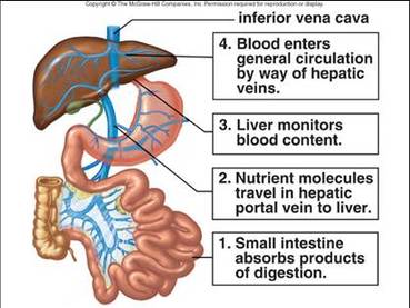
Fig.: Hepatic Portal Circulation
Coronary Circulation
This circulation supplies the heart muscle itself with oxygen and nutrients and conveys carbon dioxide and other waste products away from the heart. Two coronary arteries lead from the aorta to the heart wall, where they branch off and enter the heart muscle. The blood is returned from the heart muscle to the right atrium through the coronary vein, which enters the right atrium through the coronary sinus.
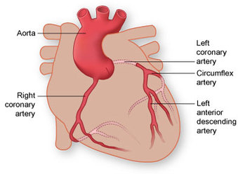
Fig.: Coronary Circulation
Valves Of The Heart
When each chamber contracts, the valve at its exit opens. When it is finished contracting, the valve closes so that blood does not flow backwards.
Valves Of The Heart
When each chamber contracts, the valve at its exit opens. When it is finished contracting, the valve closes so that blood does not flow backwards.
- The tricuspid valve is at the exit of the right atrium.
- The pulmonary valve is at the exit of the right ventricle.
- The mitral valve is at the exit of the left atrium.
- The aortic valve is at the exit of the left ventricle.
Fig.: Valves of Mammalian Heart
Mitral Valve
It prevents blood flowing from left ventricle into left atrium.
Tricuspid Valve
It allows blood to flow from right atrium into the right ventricle during diastol.
Pulmonary Valve
It prevents backflow blood into right ventricle.
Aortic Valve
It prevents blood flowing from the aorta into the left ventricle during ventricle systole.
Veins Valves
Veins also have valves which prevent backflow and maintain the direction of blood flow to heart.
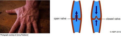
Fig.: Veins Valves
Blood Vessels
It is the main artery leaving the heart carrying oxygenated blood.
Pulmonary Artery
Pulmonaryartery: It is the only artery that carries deoxygenated blood to lungs.
Arteries
They are blood vessels that carry blood away from heart. Their walls contain elastic fibers to give characteristic elasticity.
Veins
They carry blood to the heart. They less in elastic fibers.
Pulmonary Veins
They are the only veins that carry oxygenated blood to the heart.
Blood Capillaries
They thin walled vessels in which gas exchange occurs.
Blood Vessels
- Aorta
- Arteries
- Veins
- Blood
- Capillaries
It is the main artery leaving the heart carrying oxygenated blood.
Pulmonary Artery
Pulmonaryartery: It is the only artery that carries deoxygenated blood to lungs.
Arteries
They are blood vessels that carry blood away from heart. Their walls contain elastic fibers to give characteristic elasticity.
Veins
They carry blood to the heart. They less in elastic fibers.
Pulmonary Veins
They are the only veins that carry oxygenated blood to the heart.
Blood Capillaries
They thin walled vessels in which gas exchange occurs.
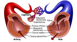
Fig.: artery, Vein and blood Capillaries
Control Of Blood Vessels
There are two main hypotheses, the Myogenic and the Metabolic.
Normal Heart Sound
With the aid of a stethoscope you can hear the characteristic sounds of the normal heartbeat, typically
described as a "lub-dub." Sometimes, a third heart sound can be heard in young. This sound is produced by the very rapid influx of blood into the ventricle. It is typically very faint, it is difficult to hear.
First Heart Tone S1(Lub)
It is caused by the closure of the atrioventricular valves and mitral at the beginning of ventricular systole.
Second Heart Tone S2 (Dub)
It is caused by the closure of the aortic valve and pulmonic valve at the end of ventricular systole.
Control Of Blood Vessels
There are two main hypotheses, the Myogenic and the Metabolic.
- Myogenic hypothesis as constant changes of stretch of the smooth muscle to maintain a constant tone.
- Metabolic hypothesis, suggests that the tone is dependant on the vasodilator substances released from
metabolism such as adenosine, lactic acid, low pH, high PCO2 and low PO2. - vasoconstrictor metabolites as serotonin released from platelets, drop in temperature, prostaglandins, Thromboxane
A2.
Normal Heart Sound
With the aid of a stethoscope you can hear the characteristic sounds of the normal heartbeat, typically
described as a "lub-dub." Sometimes, a third heart sound can be heard in young. This sound is produced by the very rapid influx of blood into the ventricle. It is typically very faint, it is difficult to hear.
First Heart Tone S1(Lub)
It is caused by the closure of the atrioventricular valves and mitral at the beginning of ventricular systole.
Second Heart Tone S2 (Dub)
It is caused by the closure of the aortic valve and pulmonic valve at the end of ventricular systole.
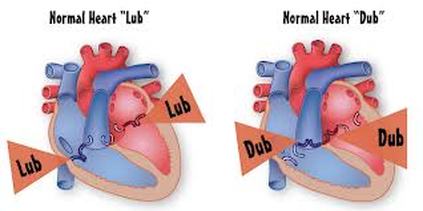
Fig.: Normal Heart Sounds
In another words, When a healthy heart pumps blood, it makes a “lub-dub” sound. The “lub” sound is the first heart sound (S1) and is caused by the closing of the mitral valve and tricuspid valve. The “dub” sound is the second heart sound (S2) and is caused by the closing of the aortic valve and pulmonary valve.
Propagation Of Heart Rate
It originates from sino atrial node (SA) in the right atrium (pacemaker). The pacemaker cells can also be controlled by the nervous system. The sympathetic nervous system can speed up the heart rate, and the parasympathetic nervous system can slow down the heart rate.
These signals spread to atrioventricular node (AV).Atrioventricle node fire action potential through bundle of His and Purkinjie
fibres causing ventricles to contract.

Fig.: Propagation Of Heart Rate
The Electrocardiogram (ECG)
ECG measures changes in electrical potential across the heart, and can detect the contraction pulses that pass over
the surface of the heart.
Recording Of ECG (Einthoven's Traingle)
- ECG is a recording of the electrical activity of the heart make from electrodes placed on the surface of the skin.
- Simply by placing the electrodes on the surface of the skin, we can find out what the heart is doing. This is because we are basically filled with NaCl and that electrolyte transfers electricity well.
- The Einthoven’s triangle set up is used to measure cardiac events. The leads consist of one positive lead, one negative lead
and one ground lead. They are placed on your right arm, left arm and left leg.
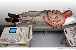
Fig.: ECG Connection
An ECG shows the sum of the electrical potentials generated by all the cells of the heart.
An ECG shows the sum of the electrical potentials generated by all the cells of the heart.
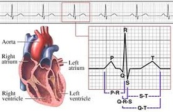
Fig.: ECG
- The P wave corresponds to the depolarization of the atrium.
- The QRS complex corresponds to the progressive way of ventricular depolarization through the heart.
- The T wave shows repolarization of the ventricles.
- Atrial repolarization is not seen because the atrium repolarizes while the ventricle is depolarizing and is incorporated into the QRS complex.
- Both the P wave and the QRS complex represent depolarisation waves.
- Subsequent to ventricular contraction, the ventricles repolarise, and the electrical currents that are produced generate the T wave.
- Therefore, the T wave is one of repolarisation and typically occurs 0.25 to 0.35 second following ventricular depolarisation.
- The interval between the beginning of the P wave and the beginning of the QRS complex is designated by the PQ interval. Repolarisation of the atria is also masked by the QRS complex.
- Since the Q wave is often absent, it is also termed the PR interval and represents the duration of time (normally about 0.16 second) between the onset of atrial contraction and the onset of ventricular contraction.
